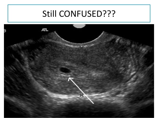
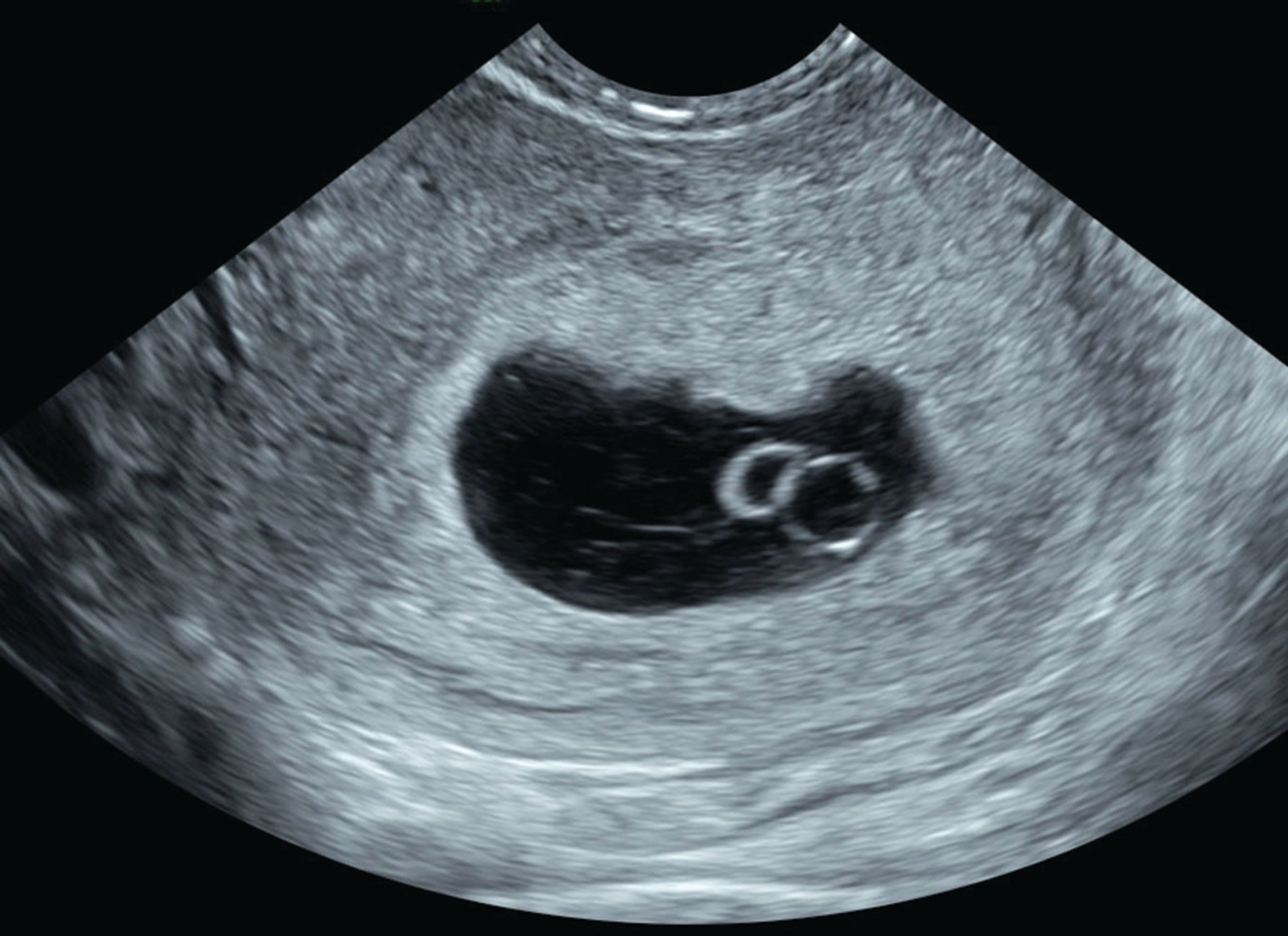
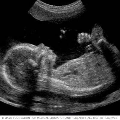
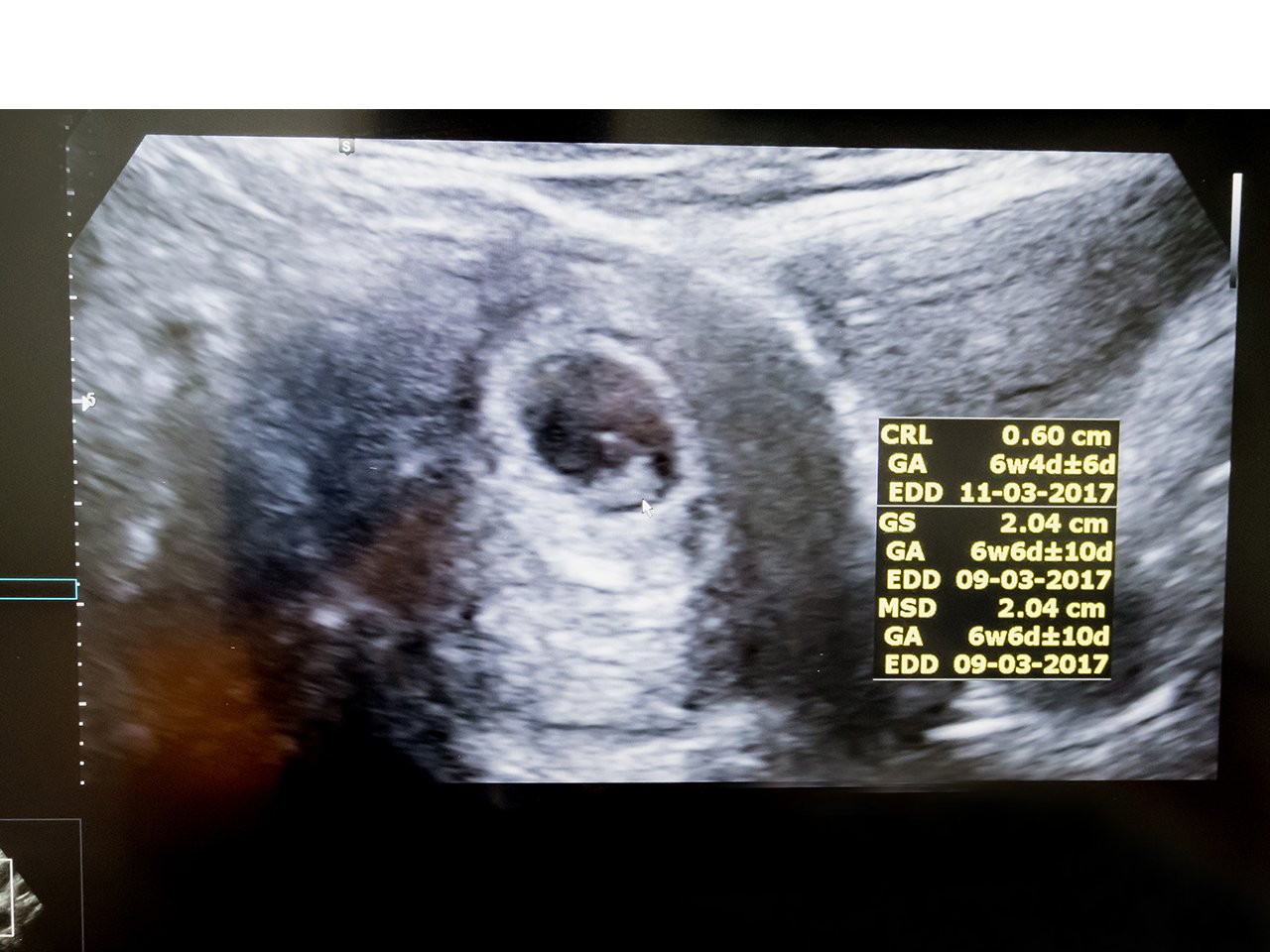
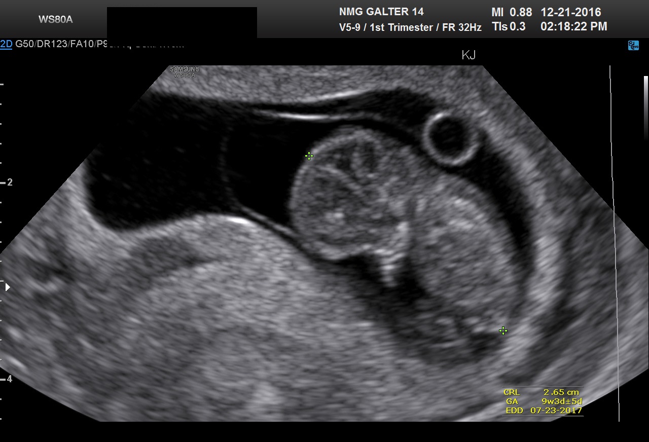
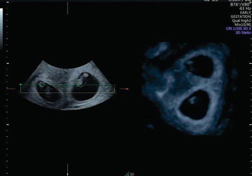
normal first trimester ultrasound sorted by
relevance

Admin16.07.2021

The 11

At 13 weeks, there was enlargement of the bladder 30 mm and bilateral hydronephrosis with reduced amniotic fluid; the pregnancy was terminated.
8003
Admin04.08.2021

Down Syndrome Markers: What Do They Mean for My Baby If They're Picked Up in an Ultrasound?

reported that the diameters of the two ventricles were similar and increased linearly with gestation from about 1.
7409
Admin16.08.2021

Normal 15 week baby Ultrasound.

There are several case reports on the sonographic diagnosis of cardiac defects at 11—14 weeks of gestation.
6705
Admin02.07.2021

Ultrasound monitoring in first trimester of pregnancy

Mild hydronephrosis or pyelectasia may be due to relaxation of smooth muscle of the urinary tract by the high levels of circulating maternal hormones, or maternal—fetal overhydration.
8506
Admin26.07.2021

First Trimester Ultrasound

At 8—10 weeks, of gestation, all fetuses demonstrate herniation of the midgut that is visualized as a hyperechogenic mass in the base of the umbilical cord; retraction into the abdominal cavity occurs at 10—12 weeks and it is completed by 11 weeks and 5 days 11,67,68.
307
Admin24.08.2021

First Trimester Ultrasound

Johnson P, Sharland G, Maxwell D, Allan L.
6203
Admin22.08.2021

First trimester screening

Table 1 - Case reports on the prenatal diagnosis of anencephaly at 11—14 weeks of gestation Author normal normal acrania anencephaly Goldstein et al.
8609
Admin30.06.2021

First Trimester Ultrasound

Diagram of early gestational sac.
1803
Admin20.08.2021

Introduction to OB Sonography: The First Trimester

It is imperative to ask your health care provider if you need to keep the bladder full before the scan.
8708
Admin09.08.2021

First trimester

Five fetuses with cardiac defects were identified, including one with a small left ventricle and pericardial effusion at 11 weeks, one with ventricular septal defect, dilated left ventricle and pericardial effusion at 12 weeks that was subsequently diagnosed as tetralogy of Fallot, one with ventricular septal defect and overriding aorta at 13 weeks, one with dextrocardia at 14 weeks that was subsequently found to also have a ventricular septal defect, and another with a single atrium and single ventricle at 14 weeks.
9603
Admin07.07.2021

The 11

In contrast, Blaas et al.
Admin22.08.2021

Ultrasound of the Normal and Failed First

The future foramina of Monro are wide during week 7.
7703